Diagram Of Liver And Blood Vessels : Arteries Of The Body Picture Anatomy Definition More - Portal, sublobular, and superficial lymphatic vessels.. They also transport blood that has been drained from the colon, pancreas, small intestine, and the stomach. The hepatic portal system is a series of veins that carry blood from the capillaries of the stomach, intestine, spleen, and pancreas to capillaries in the liver. The lymphatic vessels in the coronary ligament drain into those along the inferior vena cava. The periportal area is highly complex and consists of a dense matrix containing collagen where It is suggested that 80 % or more …
Other terms for a liver hemangioma are hepatic hemangioma and cavernous hemangioma. A liver hemangioma is made up of a tangle of blood vessels. The periportal area is highly complex and consists of a dense matrix containing collagen where The liver is supplied by two main blood vessels on its right lobe: The diagram given below represents the liver, kidney and some associated blood vessels.
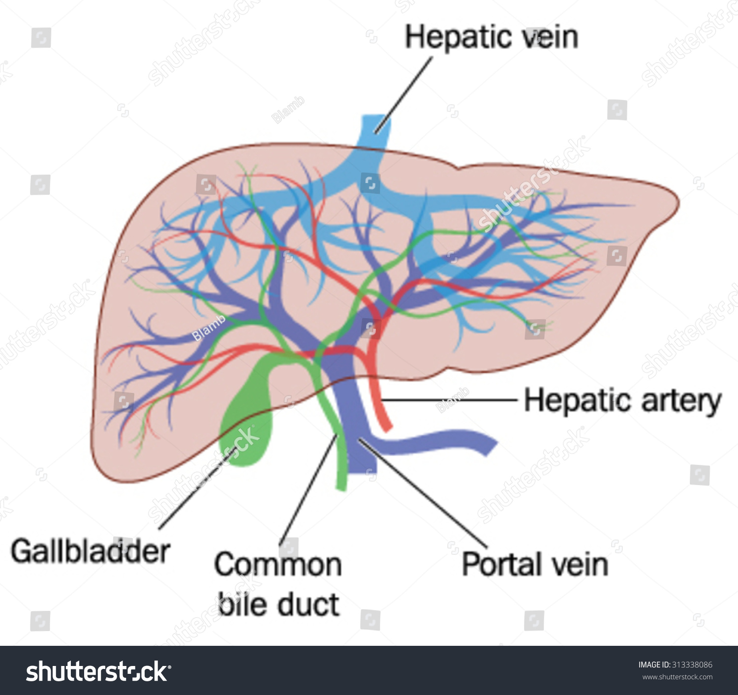
Drawing Liver Showing Major Blood Vessels Stock Vector Royalty Free 313338086 from image.shutterstock.com The hepatic portal system is a series of veins that carry blood from the capillaries of the stomach, intestine, spleen, and pancreas to capillaries in the liver. The lymphatic vessels from the lateral area of the liver convexity advance in the triangular ligament toward the diaphragm and lead into the pancreaticolienal lymph nodes. Other terms for a liver hemangioma are hepatic hemangioma and cavernous hemangioma. The lymphatic vessels in the coronary ligament drain into those along the inferior vena cava. A vessel located in the abdominal cavity that is formed by the union of the superior mesenteric and splenic veins that channel blood from the gastrointestinal tract and spleen to the capillary beds in the liver.; Blood supply of the liver and gallbladder: The hepatic lymphatic system falls into three categories depending on their locations: The liver produces a large amount of lymph, which is estimated to be 25 to 50 % of lymph flowing through the thoracic duct.
The liver is unique among organs in that it receives blood via two distinct circulatory routes:
Structure of blood vessel walls • the walls of all blood vessels, except small capillaries, have. Blood vessel (vascular) disorders of the liver usually result from inadequate blood flow—whether into or out of the liver. It is approximately three to four inches in length and is usually formed by the merging of the superior mesenteric and splenic veins behind the upper edge of the head of the pancreas. The liver is the first site of passage for venous blood arriving from the intestines via vena porta 10. It is derived from the coeliac trunk. You will also find the branch of the portal vein (distributing vein), right and left hepatic ducts (bile ducts), right and left hepatic arteries, the portal vein as well. Each of these routes provides blood of differing compositions that allow the liver to perform its unique and vital digestive and metabolic functions. Liver blood supply diagram in this image, you will find blood supply to liver, central vein system, hepatic vein, aorta, branch of hepatic artery, branch of the bile duct in it. The opposition to blood flow due to friction between blood and blood vessel walls. The areas around the influx blood vessels are named periportal. Arterioles distribute blood to capillary beds, the sites of exchange with the body tissues. The blood vessels are an intricate network of hollow tubular structures carrying blood throughout the body. They transport blood cells, nutrients and oxygen and carry away carbon dioxide and waste materials from the tissues and organs.
The portal vein brings venous blood from the spleen, pancreas, and small intestine so that the liver can process the nutrients and byproducts of food digestion. If the problem is blood flow out of the liver, blood backs up in the liver, causing congestion which can result in an enlarged liver.in either case, liver cells do not receive enough blood (called ischemia) and thus are deprived of oxygen and nutrients. A vessel located in the abdominal cavity that is formed by the union of the superior mesenteric and splenic veins that channel blood from the gastrointestinal tract and spleen to the capillary beds in the liver.; The superficial lymphatic vessels from the concave part of the liver curvature. Portal, sublobular, and superficial lymphatic vessels.
Hepatic Portal Vein Preview Human Anatomy Kenhub Youtube from i.ytimg.com Portal, sublobular, and superficial lymphatic vessels. Arteries, arterioles, capillaries, venules and veins. The hepatic lymph derives primarily from the hepatic sinusoids, and to a lesser extent from the. Tried, tested, trusted and affordable for all qpcr needs. Hepatocytes carry out many metabolic functions, including the production of bile. Structure of blood vessel walls • the walls of all blood vessels, except small capillaries, have. Kupffer cells line the liver's vascular system; • found in the liver, bone marrow, lymphoid tissue, and in some endocrine organs • allow large molecules (proteins and blood cells) to pass
They also transport blood that has been drained from the colon, pancreas, small intestine, and the stomach.
Liver blood supply diagram in this image, you will find blood supply to liver, central vein system, hepatic vein, aorta, branch of hepatic artery, branch of the bile duct in it. The hepatic lymphatic system falls into three categories depending on their locations: The hepatic lymph derives primarily from the hepatic sinusoids, and to a lesser extent from the. Blood vessels effect of liver a 2 to 1 glucose added b 2 to 1 urea removed c 3 to 1 glucose added d 3 to 1 urea removed Capillaries lead back to small vessels known as venules that flow into the larger veins and eventually back to the heart. Arterioles distribute blood to capillary beds, the sites of exchange with the body tissues. Structure of blood vessel walls • the walls of all blood vessels, except small capillaries, have. • found in the liver, bone marrow, lymphoid tissue, and in some endocrine organs • allow large molecules (proteins and blood cells) to pass If the problem is blood flow out of the liver, blood backs up in the liver, causing congestion which can result in an enlarged liver.in either case, liver cells do not receive enough blood (called ischemia) and thus are deprived of oxygen and nutrients. Blood supply of the liver and gallbladder: It is suggested that 80 % or more … The blood vessels are an intricate network of hollow tubular structures carrying blood throughout the body. Blood vessel (vascular) disorders of the liver usually result from inadequate blood flow—whether into or out of the liver.
Blood vessel (vascular) disorders of the liver usually result from inadequate blood flow—whether into or out of the liver. Tried, tested, trusted and affordable for all qpcr needs. It is part of the body's. What determines the degree of vascular resistance? The portal vein brings venous blood from the spleen, pancreas, and small intestine so that the liver can process the nutrients and byproducts of food digestion.
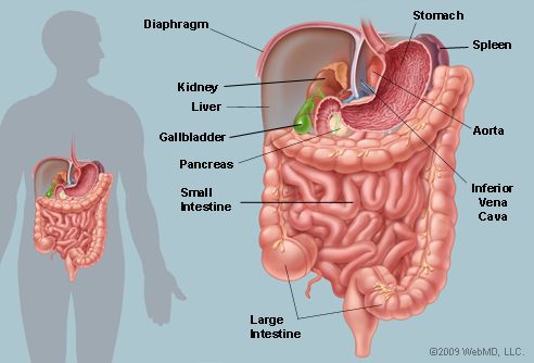
The Abdomen Human Anatomy Picture Function Parts Definition And More from img.webmd.com Arterioles distribute blood to capillary beds, the sites of exchange with the body tissues. The major one is via the hepatic portal vein (75%), which carries venous blood from the intestines , pancreas and spleen. Liver blood supply diagram in this image, you will find blood supply to liver, central vein system, hepatic vein, aorta, branch of hepatic artery, branch of the bile duct in it. Arteries, arterioles, capillaries, venules and veins. Most cases of liver hemangiomas are discovered during a test or procedure for some other condition. The heart beats continuously, pumping the equivalent of more than 14,000 litres of blood every day through five main types of blood vessels: The portal vein brings venous blood from the spleen, pancreas, and small intestine so that the liver can process the nutrients and byproducts of food digestion. Small intestine liver 12 3 after a meal, how is the blood affected by the liver as it passes between these blood vessels?
Arterioles distribute blood to capillary beds, the sites of exchange with the body tissues.
Blood vessels effect of liver a 2 to 1 glucose added b 2 to 1 urea removed c 3 to 1 glucose added d 3 to 1 urea removed They play a role in blood formation and the destruction of cellular debris. A liver hemangioma is made up of a tangle of blood vessels. Other terms for a liver hemangioma are hepatic hemangioma and cavernous hemangioma. The liver has a unique dual blood supply: The liver, as an organ, receives blood from two different sources. Liver cells, or hepatocytes, have direct access to the liver's blood supply through small capillaries called sinusoids. Tried, tested, trusted and affordable for all qpcr needs. A blood vessel that supplies oxygenated blood to the liver.; A substance, especially a coenzyme or a metal, that must be. Its normal blood volume, including both that in the hepatic veins and that in the hepatic sinuses, is about 450 milliliters, or almost 10 percent of the body's total blood volume. Kupffer cells line the liver's vascular system; The blood vessels are an intricate network of hollow tubular structures carrying blood throughout the body.
Kupffer cells line the liver's vascular system; diagram of liver. Blood supply of the liver and gallbladder:


0 Komentar